We use cookies to offer you the best experience on our site. You can find out more about the cookies we use or disable them in the settings. Cookie settings
AIDA Image Analysis Software
The AIDA Image Analyzer is designed for fast and reliable acquisition of quantitative and qualitative data from all kinds of experiments in life sciences.
The software was designed for image analysis in most applications based on image plate technology.
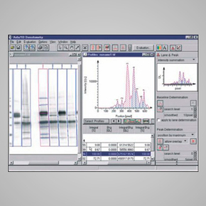
To address the needs of the different applications different modules are available.
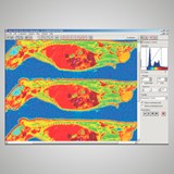
The AIDA Whole Body Autoradiography module (henceforth WBA) combines the full functionality of the AIDA 2D Densitometry module with special features required for working under GxP (Good Laboratory/Manufactory Practice) conditions. It has been primary designed for the evaluation of full body autoradiography. Since autoradiography is a 2D application, the 2D features of the WBA module are identical to the ones shipped with the 2D Densitometry module. Autoradiographs of tissue cuts result in digital images, where the signal intensity of certain areas of these images need to be evaluated. To enable quantification, these areas must be marked by an enclosure of the appropriate shape (rectangle, circle, free drawn object). For the AIDA 2D Densitometry module, the WBA module is used to evaluate the intensity within two-dimensional shapes (called regions). Tissue sections with very little radioactivity are not or hardly detected in radio-luminography. To resolve this difficulty in the WBA Professional module a visible picture of the section can be overlaid transparently over the measurement and the regions can be defined on the visible picture of the section. This option is not available in the WBA Classic edition.
Available modules: 1D Evaluation, 1D TLC; 2D Densitometry, 2D TLC; Multi Labelling, Whole Body Autoradiography, Array Metrix.


